Home
Uncategories
Loculated Pleural Effusion / Pleural Effusion - Both membranes, the visceral and parietal layer, produce and reabsorb fluid at a specific rate.
Loculated Pleural Effusion / Pleural Effusion - Both membranes, the visceral and parietal layer, produce and reabsorb fluid at a specific rate.
Loculated Pleural Effusion / Pleural Effusion - Both membranes, the visceral and parietal layer, produce and reabsorb fluid at a specific rate.. Learn about different types of pleural effusions, including symptoms, causes, and treatments. Slowly clearing infections in the pleural space are a source of substantial morbidity. Loculated pleural effusion must be included in the differential diagnosis of roentgenographic densities in the chest when seen in subcostal as well as in interlobar locations. Pleural effusion can be resolved by putting a pleural drain, performing pleurodesis, vats, or thoracotomy. 1 article features images from this case 20 public playlist includes this case
Find symptoms,causes and treatments of pleurisy.for your health. The pleural fluid is called a transudate if it permeates (transudes) into the pleural cavity through the walls of intact pulmonary vessels. Nursing care plan 3 nursing diagnosis: The lack of specificity is mainly due to the limitations of the imaging modality. Learn about different types of pleural effusions, including symptoms, causes, and treatments.
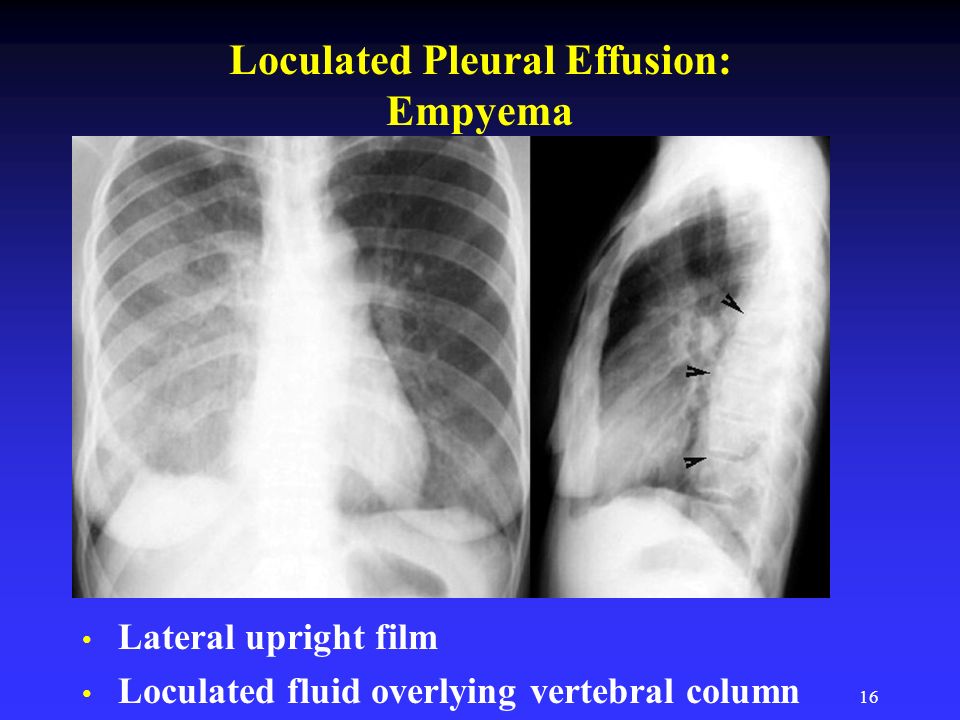
Icu patients cannot sit up and the effusion layers posteriorly.
(2) the gram stain or culture is positive; Surgical thoracostomy tube placement and radiologically guided catheter drainage are standard therapy for loculated pleural fluid collections. Causes of an exudative effusion are malignancy, infection, or inflammatory disorders such as rheumatoid arthritis. Icu patients cannot sit up and the effusion layers posteriorly. Pleural effusions in the intensive care setting. Search for loculated pleural effusion. The lack of specificity is mainly due to the limitations of the imaging modality. Slowly clearing infections in the pleural space are a source of substantial morbidity. Pleural effusion is the accumulation of excess fluid in the lung space, the space between the membrane lining the lungs and the membrane lining the chest wall. Cytological analysis of pleural fluid showed a negative result for malignant tumor cells. Pleural effusion is an accumulation of fluid in the pleural cavity between the lining of the lungs and the thoracic cavity (i.e., the visceral and parietal pleurae). Loculated pleural effusion must be included in the differential diagnosis of roentgenographic densities in the chest when seen in subcostal as well as in interlobar locations. Find symptoms,causes and treatments of pleurisy.for your health.
(2) the gram stain or culture is positive; A loculated pleural effusion are most often caused by an exudative (inflammatory) effusion. The category 3 effusion meets at least one of the following criteria: Sometimes in the setting of pleuritis, loculation of fluid may occur within the fissures or between the pleural layers (visceral and parietal). The lack of specificity is mainly due to the limitations of the imaging modality.
3 from Learn about different types of pleural effusions, including symptoms, causes, and treatments. Cultures of pleural fluid and blood showed no growth of aerobic or anaerobic organisms. Normally, a small amount of fluid is present in the pleura. Enlarged mediastinal lymph nodes, possibly reactive. (2) the gram stain or culture is positive; Loculated pleural effusion (427895005) recent clinical studies. Causes of an exudative effusion are malignancy, infection, or inflammatory disorders such as rheumatoid arthritis. Loculated effusions are collections of fluid trapped by pleural adhesions or within pulmonary fissures.
Pleural effusion that is confined to one or more fixed pockets in the pleural space.
Loculated Transudative Pleural Effusion Masquerading As Right Upper Lobe Consolidation In A Haemodialysis Patient Kho Ame Case Reports from cdn.amegroups.cn A right loculated pleural effusion is still evident. Surgical thoracostomy tube placement and radiologically guided catheter drainage are standard therapy for loculated pleural fluid collections. Loculated right pleural effusion with foci of atelectasis and consolidative changes concerning for pneumonia. Loculated pleural effusion masquerading as mediastinal tumour had been reported but pleural effusion that conformed to the contour of a lung lobe is rare. 1 article features images from this case 20 public playlist includes this case Search for loculated pleural effusion. Cytological analysis of pleural fluid showed a negative result for malignant tumor cells. Nursing care plan 3 nursing diagnosis:

0 Comments:
Posting Komentar